Early detection saves lives. Tissue AI empowers your practice to identify oral health concerns before they become serious problems.
What is Tissue AI?
Tissue AI is scanO's breakthrough technology that analyzes soft tissue in the mouth using simple photographs. Whether captured with your scanO Air device or mobile app, our AI instantly evaluates oral tissue to help you provide better patient care.
The result? Faster diagnosis, earlier intervention, and peace of mind for both you and your patients.
What Tissue AI Detects
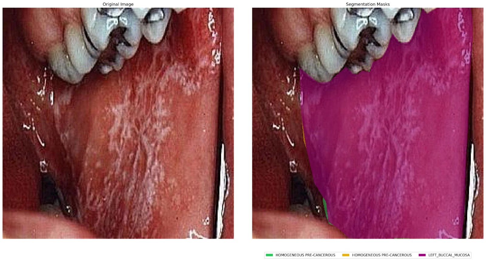
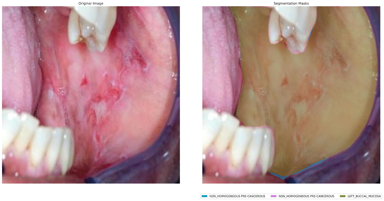
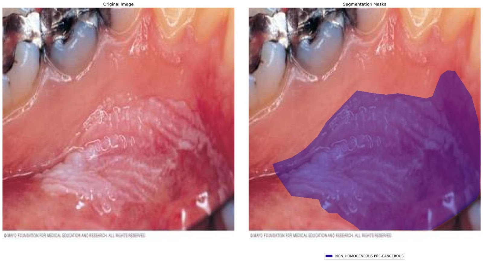
Visual Assessment Categories
Homogeneous Lesions
Uniform appearance and texture
Consistent coloring throughout
Even surface patterns
Non-Homogeneous Lesions
Mixed textures and appearances
Varied coloring or patterns
Irregular surface characteristics
Ulcerative Lesions
Open sores or wounds
Tissue breakdown
Healing assessment
Clinical Risk Categories
Non-Cancerous Conditions
Benign tissue changes
Common oral variations
Low-risk findings
Pre-Cancerous Conditions
Early warning signs
Tissue changes requiring monitoring
Preventive care opportunities
Cancerous Conditions
Malignant tissue identification
Urgent referral recommendations
Critical early detection
Key Detection Markers
Tissue AI analyzes multiple visual indicators simultaneously:
Color Changes - Unusual redness, whiteness, or discoloration
Texture Variations - Smooth, rough, or bumpy surfaces
Growth Patterns - Abnormal tissue development
Border Definition - Clear vs. irregular edges
Tissue Hardening - Areas of unusual firmness
Surrounding Impact - Effects on nearby healthy tissue
How We Made It Possible – Just From An Image
The Power of Visual Intelligence
What makes Tissue AI remarkable is its ability to extract comprehensive screening insights from pictures taken in a guided manner. Here's how we transformed simple imaging into powerful clinical intelligence:
Advanced ML model:
Trained to recognize subtle visual patterns indicating different tissue conditions
Analyzes thousands of microscopic details in each image that might be missed by the human eye
Identifies relationships between color, texture, and shape that reveal tissue health status
Comprehensive Visual Analysis Every image is evaluated across multiple dimensions simultaneously:
Surface Characteristics: Smooth, rough, irregular, or ulcerated appearances
Color Mapping: Precise analysis of color variations and their clinical significance
Border Analysis: Sharp, diffuse, or irregular boundary patterns
Texture Assessment: From homogeneous surfaces to complex mixed patterns
Contextual Understanding: How lesions relate to surrounding healthy tissue
Real-World Training
Trained on thousands of real patient images from diverse clinical settings
Professionally annotated by experienced dental experts
Continuous learning from new cases to improve accuracy over time
Quality-controlled dataset ensures only the clearest, most relevant images contribute to learning
Smart Clinical Correlation Tissue AI doesn't just see—it understands. The system correlates visual findings with:
Known risk factors and patient habits
Common presentation patterns for different conditions
Clinical significance of various combinations of visual markers
Appropriate urgency levels for different findings
The Result: A single photograph becomes a comprehensive tissue health assessment, providing insights needed for confident clinical decision-making.
Risk-Based Screening Approach
Risk Scores & Probability Analysis
Risk Scores: Easy-to-understand ratings indicating the likelihood of concerning findings
Probability Scores: Precise percentages showing the confidence level for each assessment
Priority Flagging: Immediate identification of patients who need closer attention or specialist referral
Screening Tool, Not Diagnosis
Helps identify patients who may need further investigation
Supports clinical judgment with data-driven insights
Flags high-risk cases that might otherwise be missed during routine care
Provides consistent screening standards across your entire practice
Finding High-Risk Patients
Catch elevated-risk patients before symptoms become obvious
Ensure no high-risk case goes unnoticed during busy clinic days
Provide objective screening criteria to complement clinical experience
Create a safety net for comprehensive patient care
Proven Performance
Current Capabilities
75% Screening Accuracy – Strong performance with continuous improvement
Expert-Validated – All training data reviewed by dental professionals
Quality-Controlled – Only high-resolution, clinically relevant images used
Track Record of Excellence Our companion technology, Tooth AI for hard tissue screening, already achieves 96% accuracy. Tissue AI follows the same proven development path.
Transform Your Practice
For Your Team
Faster Decisions – Instant preliminary assessment
Enhanced Confidence – AI-powered second opinion
Better Documentation – Clear visual evidence for records
Streamlined Workflow – Seamless integration with existing processes
For Your Patients
Earlier Detection – Catch problems before they advance
Clear Communication – Visual explanations they can understand
Faster Treatment – Quicker path to appropriate care
Peace of Mind – Thorough, technology-assisted examination
Real-World Application
Simple Process:
Capture image with scanO engage
Tissue AI analyzes the image instantly
Receive categorized assessment with visual indicators
Make informed treatment decisions
Communicate findings clearly to patients
Immediate Benefits:
Spot potential issues during routine checkups
Provide evidence-based referrals when needed
Track changes in tissue over time
Enhance patient education and engagement
The Future is Now
Tissue AI isn't a prototype or concept—it’s actively working in dental practices today. As more practitioners use the system, it becomes smarter and more accurate, benefiting the entire dental community.
Your practice doesn't just use advanced AI—it helps advance the field.
Ready to Enhance Your Practice?
Join the growing community of dental professionals using Tissue AI to provide superior patient care. Early detection, better outcomes, and confident diagnosis are just a click away.
Disclaimer: Tissue AI is a screening tool designed to support clinical decision-making by identifying high-risk patients who may require further evaluation. It provides risk and probability scores to aid in diagnostic screening but does not provide definitive diagnosis. All findings should be evaluated in conjunction with comprehensive clinical examination and professional clinical judgment.
About the Author:
Reviewed By:
Subscribe to our newsletter
Thank you! Your submission has been received!
Oops! Something went wrong while submitting the form.