


Quick answer: The dentistry technology that matters most for Indian clinics in 2026 falls into three groups. Imaging: digital radiography (RVG), CBCT and intraoral cameras. Digital workflow: intraoral scanners, CAD/CAM milling and 3D printing. Intelligence and access: AI image analysis, AI-assisted screening, practice management software and teledentistry, plus dental lasers for selected procedures. Most clinics should start with digital radiography, an intraoral camera and practice software, then add scanning, CAD/CAM or AI based on case mix.
This guide explains what each tool does, what it changes in a real Indian clinic, and which clinics benefit first. It replaces our 2019 list of top technologies in dentistry, because the market has changed a great deal since then.
Two pressures push clinics toward better technology. The first is disease burden. According to the World Health Organization's oral health fact sheet (March 2025), oral diseases affect nearly 3.7 billion people. The WHO also notes that access to primary oral health services is often low because professionals and facilities are unevenly distributed.
The second is patient expectation. Patients now expect to see what the dentist sees, receive records digitally, and understand treatment before they agree to it. Technology that improves visibility, speed and communication helps a clinic meet both pressures without adding chairs.
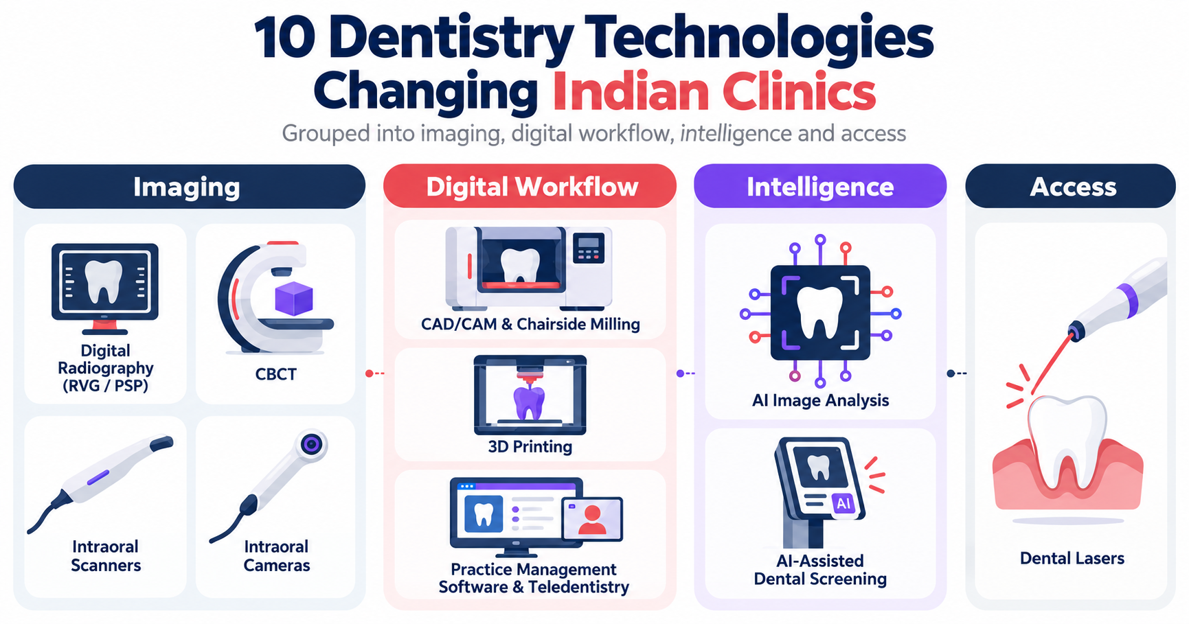
Digital intraoral sensors (commonly called RVG) and phosphor plate systems replace film. Images appear on screen within moments, can be enlarged and annotated, and are easy to store and share. For most general practices, digital radiography is the first imaging upgrade.
In India, dental X-ray equipment is regulated by the Atomic Energy Regulatory Board (AERB), and dental radiology users complete registration through AERB's e-LORA portal. For pricing, models and compliance, see our guide to dental X-ray machine price in India.
CBCT produces three-dimensional images of teeth, bone and surrounding structures. It is most valuable for implant planning, complex endodontics, impacted teeth and surgical cases. Because it is a larger investment and carries its own regulatory steps, many general practices refer out to an imaging center and buy only when implant and surgical volume justifies it.
Intraoral scanners capture a digital 3D impression of the teeth, replacing trays and impression material for many restorative, implant and aligner cases. The data goes straight to a lab or a chairside milling unit. Prices range widely by brand and bundle; our guide to intraoral scanner price in India breaks down the bands, hidden costs and ROI questions.
Computer-aided design and manufacturing lets a clinic design and mill crowns, inlays and onlays, sometimes in a single visit. The real decision is workflow: chairside milling suits high-volume restorative practices, while most clinics get similar benefits by sending digital scans to a lab with CAD/CAM capability.
Desktop 3D printers produce models, surgical guides, night guards, temporary restorations and aligner models from digital files. Printing works best in clinics that already scan digitally and have someone on the team willing to own the post-processing workflow.
An intraoral camera shows the patient a magnified view of their own teeth on a screen. It is a relatively low-cost way to make case acceptance conversations more visual. Our guide to intraoral cameras for dental clinics covers types, prices and a buying checklist.
AI software can highlight suspected caries, bone loss or other findings on radiographs and images for the dentist to review. The evidence is encouraging but still maturing. A 2025 umbrella review with meta-analysis in PLOS One reported pooled sensitivity of 0.85 and specificity of 0.90 for AI caries detection across imaging types, while cautioning that AI should not be a substitute for human judgment and that real-world validation is still needed. Clinical decisions stay with the dentist.
A newer category uses AI on intraoral photographs to screen patients for visible oral health concerns before or outside a full examination. Screening is not diagnosis: it helps flag who may need a dental visit and helps patients understand why. It is useful in reception areas, camps, schools and corporate programs. Our explainer on how AI dental screening works covers the workflow and its limits.
Soft-tissue and hard-tissue lasers are used for procedures such as gingival contouring, frenectomy, periodontal therapy and some cavity preparations. They require specific training and safety protocols. Lasers make sense when a clinic's case mix includes enough of these procedures and the dentist has completed hands-on training.
Cloud practice management software handles appointments, records, billing, imaging integration and reminders. Teledentistry adds remote consultations and follow-ups. These are the backbone that makes every other tool useful, because images and reports are only valuable if they are organized and retrievable. Any system that stores patient records must follow India's Digital Personal Data Protection Act, 2023, including consent and access controls.
Use this decision table to match tools to problems. Price columns are intentionally left out where we do not have verified 2026 figures; see the linked guides for sourced price ranges.
Stage 1: new single-chair clinic. Start with digital radiography, an intraoral camera and cloud practice software. These three improve diagnosis support, patient communication and record keeping from day one.
Stage 2: established two to three chair clinic. Add an intraoral scanner if restorative, implant or aligner cases are growing, and consider AI-assisted screening at reception to help more walk-in and recall patients understand their oral health.
Stage 3: multi-chair clinic or chain. Standardize imaging and AI analysis across branches, evaluate CBCT or a referral partnership, and consider CAD/CAM or 3D printing if case volume supports an in-house workflow.
Three directions are visible in 2026. Workflows are becoming connected, so scans, images and reports move between the chair, the lab and the patient's phone without re-entry. AI is moving from research into everyday support tools, with growing attention to validation and responsible use. And screening is moving outside the operatory, to reception areas, schools, camps and workplaces, so more people are assessed before problems become severe.
For a broader view of how AI is being applied across diagnosis support, planning and practice operations, read our overview of AI in dentistry.
In the roadmap above, AI-assisted screening belongs to Stage 2, when a clinic has steady walk-ins and recall patients but limited chair time. scanO air sits in that visual oral screening category. It uses intraoral photos to create a patient-facing report that helps walk-in and recall patients understand their oral health before they see the dentist. It is not a substitute for an intraoral scanner, radiographs, a clinical examination, diagnosis or treatment planning, and the dentist decides the next clinical step.
The most talked-about dentistry technologies in 2026 are AI image analysis and AI-assisted screening, intraoral scanning with connected lab workflows, CBCT for 3D planning, and in-clinic 3D printing. For most Indian clinics, the practical upgrades are still digital radiography, intraoral cameras and practice management software.
Most new clinics benefit first from digital radiography, an intraoral camera and cloud practice management software. Add an intraoral scanner, CBCT or CAD/CAM when your case mix and patient volume justify the investment.
Yes. AI is used in India for radiograph analysis support, AI-assisted screening from intraoral photos, and practice analytics. AI tools support the dentist's judgment; they do not diagnose on their own, and clinical decisions stay with the dental professional.
Dental X-ray equipment in India is regulated by the Atomic Energy Regulatory Board (AERB), and dental radiology users complete registration through AERB's e-LORA portal. Confirm the exact requirements for your equipment type with AERB or your supplier before installation.
No. An intraoral camera captures magnified 2D photos and video for patient communication and documentation. An intraoral scanner captures a 3D digital impression used to design restorations, aligners and implant work.
Digital dental technology covers tools that capture, store or use dental information digitally, such as digital X-rays, CBCT, intraoral scanners, CAD/CAM, 3D printing, AI software and practice management systems.
Start with the problems your clinic faces most often, then match them to the tools above. If patient understanding and early screening are high on your list, read our comparison of an intraoral scanner vs AI dental screening to see where a screening step can sit in front of your existing examination workflow.
For the product workflow, see scanO air.