

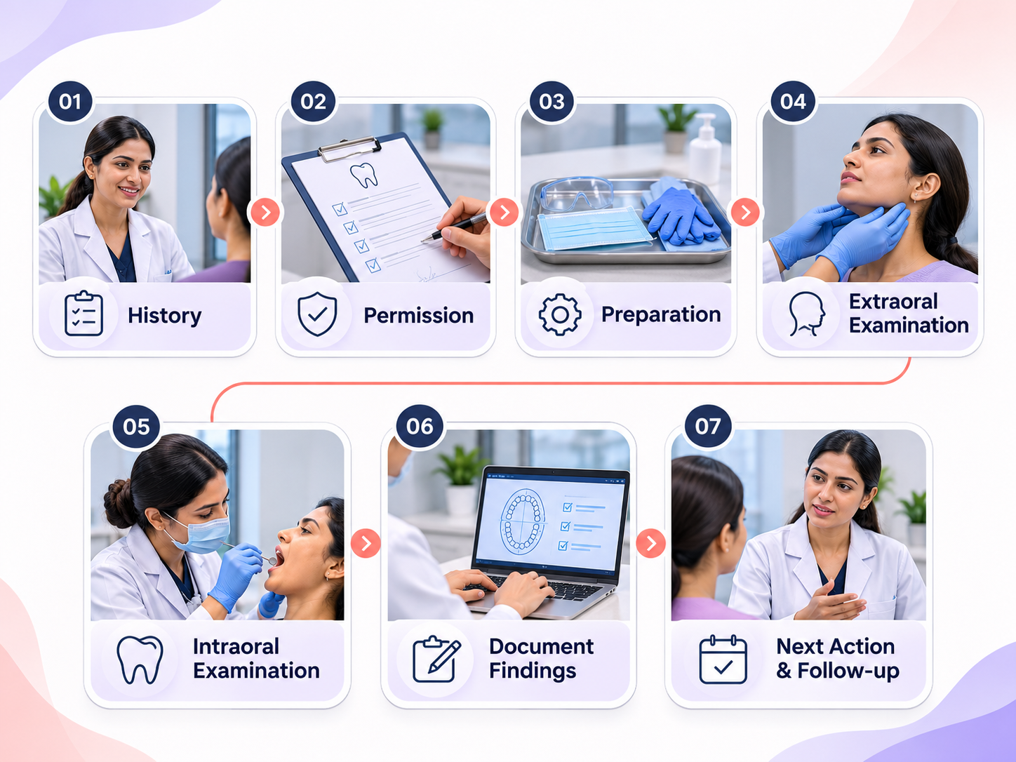
Quick answer: Oral cancer screening for a dentist starts with history and a conventional visual and tactile examination. A suspicious finding needs a documented clinical decision and prompt specialist assessment or biopsy as appropriate. Photographs support the record; they do not rule out cancer. The seven steps below organise the consultation, rather than define a validated new diagnostic test.
A patient may book for a fractured filling and never mention a change elsewhere in the mouth. A consistent examination sequence helps the team avoid letting the chief complaint become the entire consultation. It also creates a clear record of what was examined, what could not be assessed and what happens next.
A screening encounter identifies findings that need further assessment. It cannot establish histology, exclude every disease or predict an individual lesion's future. Keep the terms finding, working assessment and confirmed diagnosis separate in the notes. Do not describe a reassuring photograph as a cancer-negative certificate.
The ADA oral cancer guideline places conventional examination at the centre of assessment. Its updated guidance is particularly relevant when clinics consider adjunct technologies. Use the living guideline for current clinical recommendations rather than relying on a vendor's description of its device.
Ask about the concern, its duration and changes. Record tobacco, areca nut and alcohol exposure sensitively, together with relevant medical and dental history. Explain why an examination beyond the painful tooth is useful. Offer privacy and a chance to ask questions. Screening consent does not automatically authorise promotional photography.
Use suitable lighting, personal protective equipment and the instruments required for the clinician's examination. Explain touch before palpation. Position the patient so the clinician can examine systematically without rushing. If access, discomfort or cooperation limits the examination, record the limitation and arrange an appropriate next step.
Assess the face, lips and relevant extraoral structures as indicated by training and the patient's presentation. Document abnormalities precisely rather than recording only a generic tick. A concerning symptom or finding warrants clinical assessment even when no striking photograph can be captured.
Use the same agreed sequence for the lips, buccal mucosa, gingival and alveolar tissues, tongue, floor of mouth and palate, with other areas assessed as clinically appropriate. Combine observation with tactile examination where indicated. A sequence is a memory aid, not a substitute for professional judgment.
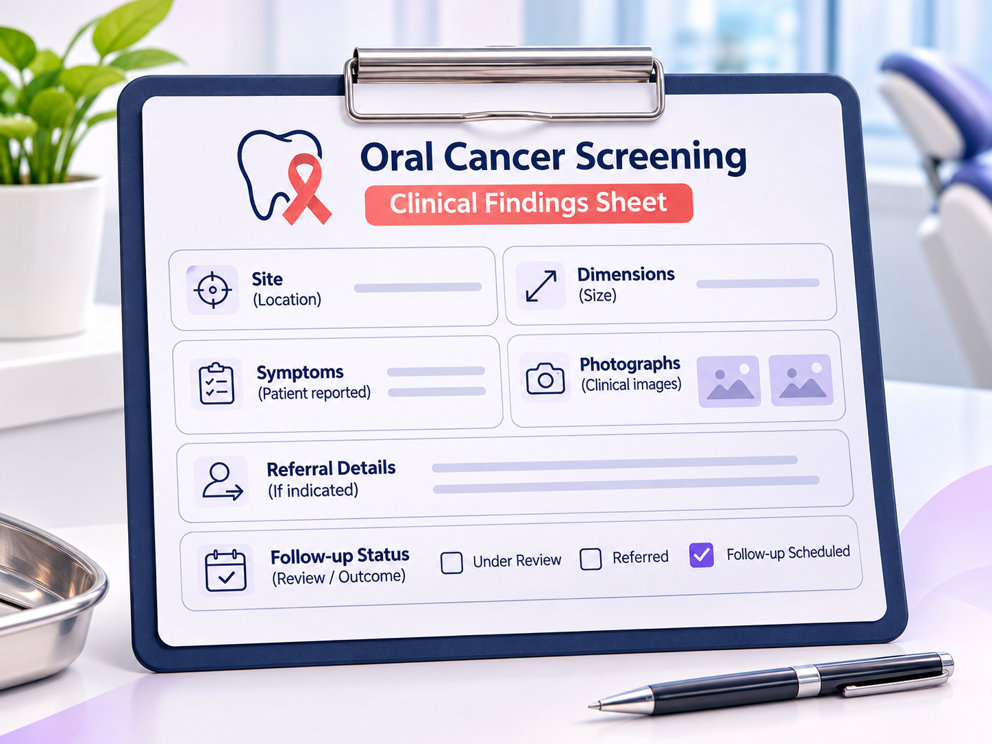
Record the site, side, dimensions where measurable, appearance, texture, symptoms and reported duration. Separate what the patient reports from what the examiner observes. A consented clinical image can help communication and comparison, but lighting, angle and image quality can change how tissue appears.
For a suspicious lesion, act on the clinical assessment without waiting for an optional device result. Name the receiving clinician or service and provide the handover. Do not use an arbitrary observation period to delay referral of a suspicious finding. If review is appropriate, document why, when and who is responsible.
Say what was seen, what remains uncertain and why the next assessment matters. Ask the patient to describe the agreed next step in their own words. Give clear contact instructions and record whether referral was accepted, declined or could not be arranged. Track attendance rather than assuming that a referral letter completes care.
Do not advertise a guaranteed examination time. The worksheet below uses planning allocations for team preparation, not clinical time limits. History, examination and counselling may take longer when a finding needs attention. A busy reception area should never determine how much time a suspicious finding receives.
Consider a fictional adult attending for sensitivity who reports a persistent change on the side of the tongue. The dentist records the separate complaint, examines the area and makes an appropriate referral. The receptionist helps arrange the appointment without discussing sensitive details in the waiting room. The tracker records the referral status, not an unconfirmed cancer diagnosis.
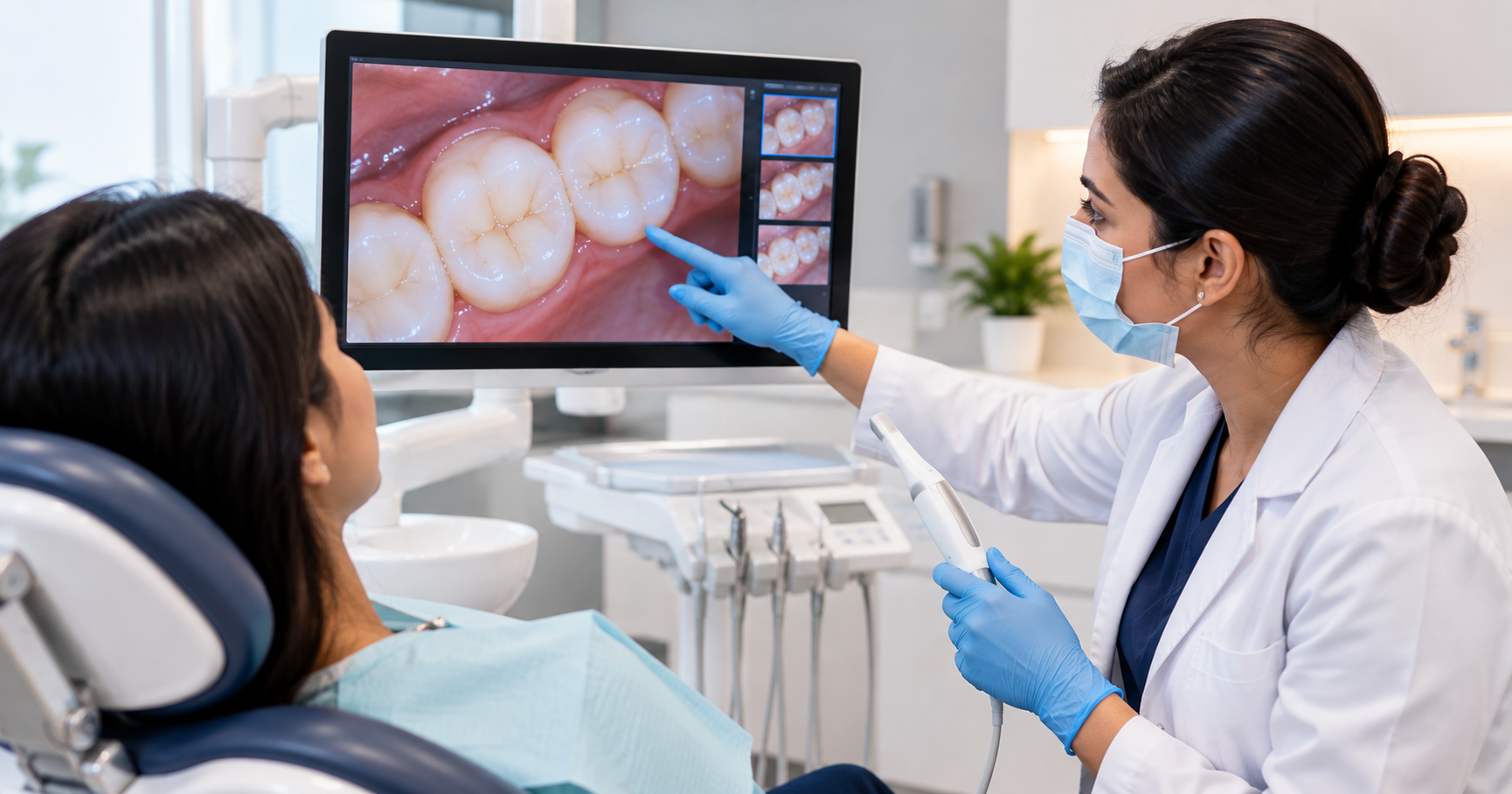
Digital records and images can make the handover more consistent. scanO air may support oral screening documentation and patient communication within its verified intended use. This article does not position it as an oral cancer detection device. Conventional examination and clinician-led decisions remain essential.
Who reviews an incomplete examination? Where are consented images stored? Who checks that a referral was received? What happens when the patient cannot afford the receiving service? Write the answers into the clinic's process so they do not depend on whichever staff member happens to be available.
Use the chairside worksheet below at the next team briefing. For the broader technology context, read how AI dental screening works.
At a team review, select a small, appropriately de-identified set of completed encounters. Ask whether another clinician could understand what happened without speaking to the original examiner. A tick saying mouth checked is less useful than a structured account of the areas assessed, the limitations and the decision. The audit should identify documentation gaps, not retrospectively invent findings that were never recorded.
Keep the audit separate from the original clinical record. If a clarification is needed, use the clinic's approved amendment process so the original entry and later clarification remain distinguishable. Do not silently edit an earlier note to make the encounter appear more complete. The aim is a trustworthy record and a better process for the next patient.
Review referral logistics as well as examination documentation. A letter can be clinically clear but operationally unusable if it lacks a receiving service or contact route. Ask the coordinator to demonstrate how they would help a patient who cannot reach the first service. The examiner should remain available for clinical questions that arise during that handover.
Patients may interpret a recommendation for further assessment as confirmation that something serious has already been found. Explain clearly what the examination established, what remains uncertain and why additional assessment is necessary. Use a calm, specific next step rather than a vague warning. Avoid offering reassurance that goes beyond the available clinical evidence.
AI-powered screening tools such as scanO Air can support dental professionals in identifying oral health concerns that may require closer examination. However, screening findings should not be presented as a confirmed diagnosis. Clinical interpretation, appropriate examination and specialist referral remain essential when potentially concerning findings arise.
For a fictional case, compare two explanations during a staff briefing. The first says the scan detected cancer. The second explains that the dentist identified an area requiring specialist assessment and provides clear instructions for arranging that appointment. Ask staff which statement overstates diagnostic certainty and which communicates an appropriate next action. This exercise reinforces responsible clinical communication without using real patient information.
Finally, confirm that the patient can access their referral information after leaving the clinic. A printed referral letter, an agreed digital communication method and a named contact may all be helpful, depending on patient preferences and local arrangements. A successful message delivery notification does not necessarily mean the patient has read, understood or acted on the referral instructions.
For further practical context, see dental camp operations. These resources support planning; adapt decisions to your clinic and review clinical requirements with qualified professionals.