


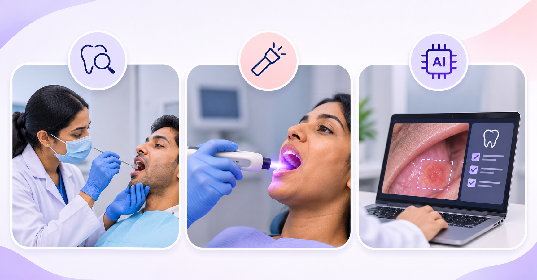
Quick answer: An oral cancer screening device cannot replace a dentist's examination or the diagnostic pathway for a suspicious finding. Compare tools by their intended use, evidence and effect on decisions, not their promotional label. The ADA's 2026 guidance advises against using light-based adjuncts to determine biopsy or referral decisions.
A product demonstration can make every abnormal area appear obvious. That does not show how the tool behaves in an ordinary clinic with inflammation, restorations, variable lighting and uncertain findings. Purchasing begins with a more useful question: what decision would this device improve, and what evidence supports that change?
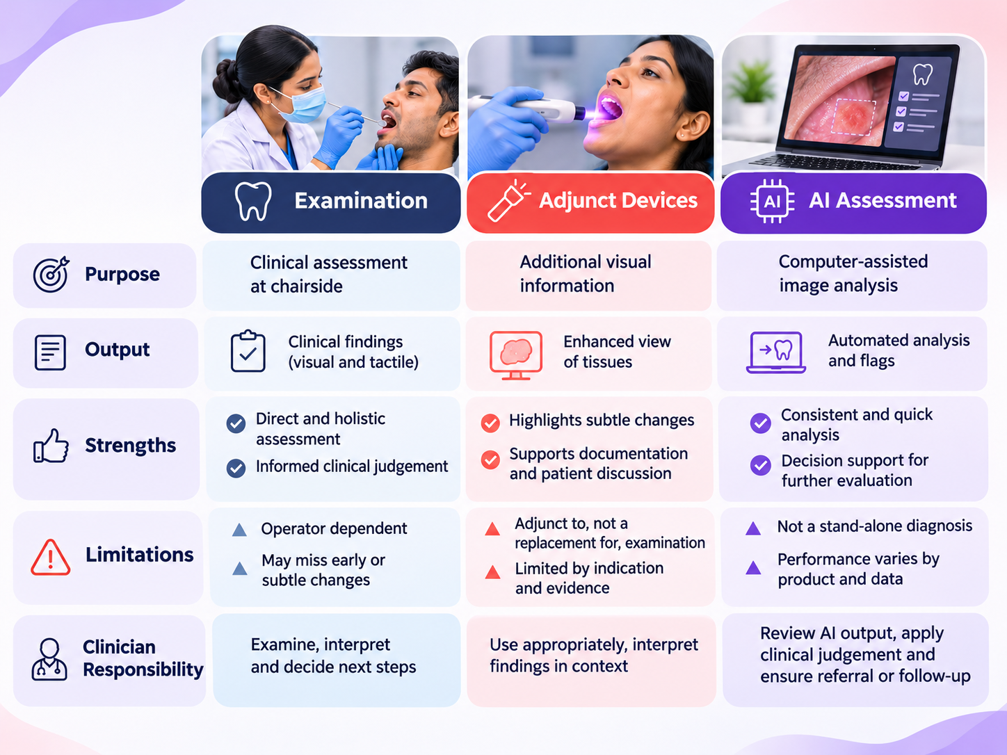
Separate three possible jobs: helping a clinician examine, recording a visible finding and determining whether further assessment is required. A camera may improve documentation without being validated for disease classification. A classification model may have research results without evidence for independent use in a community setting.
Write the intended decision into the purchase brief. If the answer is better patient communication, assess image quality, explanation and records. If the answer is lesion triage, require evidence relevant to that specific task and professional review of the proposed pathway.
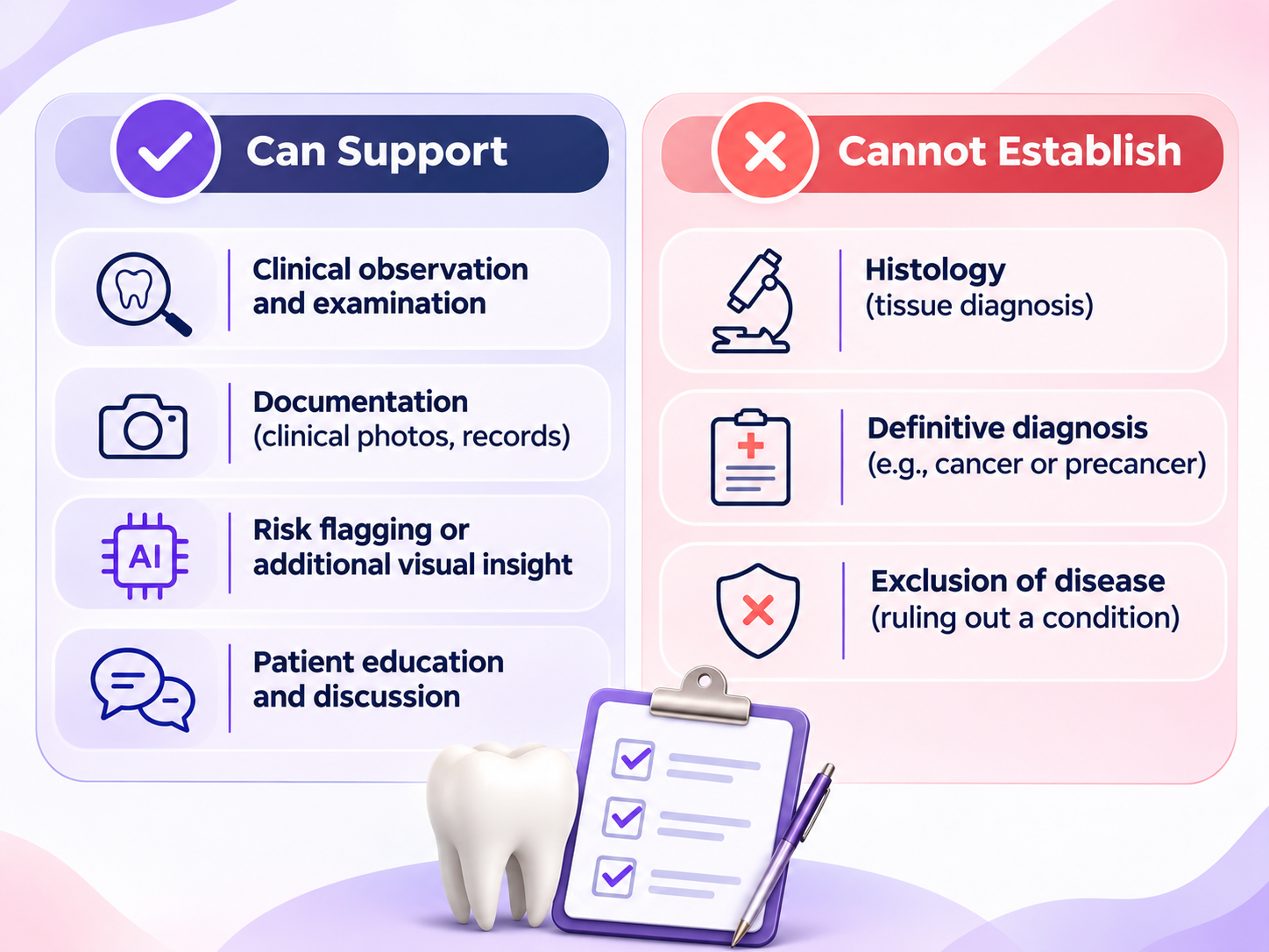
History, visual examination and tactile assessment are not interchangeable with a screen image. The dentist considers symptoms, clinical context, access limitations and the full examination. A suspicious finding should not wait for an optional adjunct to become available or produce a reassuring result.
The 2026 ADA light-based adjunct guideline gives conditional recommendations against using these tools to determine the need for biopsy or referral. Its certainty assessments matter: an attractive image is not the same as demonstrated benefit in the intended clinical decision.
Different devices may use different optical principles. Do not treat fluorescence loss or colour change as a diagnosis. Ask for the exact intended indication, the clinical population studied and the reference standard. Clarify whether the advertised performance came from a selected set of lesions or routine patients.
In the demonstration, include normal variation and common confounding conditions within an ethically appropriate evaluation. The question is not whether the image looks dramatic. It is whether the clinician receives reliable, relevant information without delaying the established pathway.
Vital staining and cytology are different adjunct categories. A recommendation about one category does not establish the value of another. Review their current guidance, sampling requirements, interpretation, costs and limitations separately. Do not combine several adjuncts into a package and call the package a definitive cancer test.
If sampling or laboratory interpretation is involved, ask who performs it, where the sample goes and how inadequate samples are handled. A clinic needs a process for an inconclusive result as well as a positive one. The purchasing team should price repeat work and referral coordination.
An AI system may analyse photographs, other optical images or histology. Those input types and clinical roles are not equivalent. Require the model version, target population, reference standard and independent validation relevant to the proposed use. A published study about another model is not evidence for the device on offer.
Ask how the system handles poor images and cases outside its validated scope. An explicit unable-to-assess result may be safer than forcing every case into a reassuring or concerning category. The operating procedure must say what the clinician does when the output conflicts with the examination.
Include training, disposable items, cleaning, software, maintenance, repeat captures and referral administration. A low purchase price can become expensive if every session needs manual rescue. Conversely, a sophisticated report may have little value if it cannot be saved to the correct patient record.
scanO air can be discussed as a screening documentation and patient-communication option. This comparison does not describe it as a cancer detection device, and no cancer sensitivity or specificity is attributed to it. Ask the product team to confirm any proposed clinical use in writing.
Use the decision table as a procurement meeting agenda. Start with the chairside examination workflow, then record which additional task, if any, remains unresolved.
Ask the supplier to show the complete operating process and describe cases the system cannot assess. Use synthetic or appropriately authorised material and make the test conditions explicit. A demonstration on selected images should be labelled as such; it does not become a clinical performance evaluation simply because the clinic watched it in person.
Before the session, write down the decisions the purchasing team must make. These could include whether the output adds useful documentation, whether a clinician can recognise an inadequate result and whether the record can be exported. Keep the evidence question separate from interface preference. A pleasant interface can improve usability without establishing diagnostic value.
Have the clinician describe what they would do without the adjunct and what, if anything, would change after seeing it. If the intended action remains the same, the buyer should understand whether the benefit is documentation, education or another non-diagnostic task. That distinction can justify a purchase more honestly than an unsupported early-detection claim.
Keep research funding, author relationships and vendor involvement visible when assessing evidence. A disclosed relationship does not automatically invalidate a study, but it is relevant context. Equally, a paper's independence does not make its findings transferable to a different commercial product. Ask whether the evaluated version and the marketed version are actually the same.
Record missing details as questions for the reviewer. A report that omits patient-level splitting, reference standards or image exclusions should not be summarised as fully validated. The purchasing team can ask for the underlying publication or protocol rather than guessing what the missing methods probably were.
Finally, separate the procurement decision from permission to expand clinical use. The clinic might accept a camera for documentation while rejecting unsupported claims about lesion classification. Write that boundary into training and patient-facing language. It should remain clear when a salesperson, staff member or later marketing agency prepares new copy about the equipment.
For further practical context, see AI dental screening workflows and dental camp operations. These resources support planning; adapt decisions to your clinic and review clinical requirements with qualified professionals.