
Many practices still treat oral cancer detection as a “visual catch” problem. If you look carefully, you will see it. That assumption breaks in real clinics because early lesions often look non-specific, lighting is inconsistent, documentation is variable, and clinician thresholds differ across chairs and sites. The result is not ignorance. It is missed escalation.
This matters because oral cancer is not rare noise in the background. Globally, lip and oral cavity cancer alone accounted for roughly 390,000 new cases and 188,000 deaths in 2022. When diagnosis shifts late, outcomes and costs compound quickly, including more complex surgery, longer rehabilitation, and higher drop-off in follow-through.
Afrtificial Intelligence in oral cancer does not “replace the exam.” It tightens consistency around what gets flagged, tracked, re-evaluated, and referred. That is where operational risk usually hides.
Assertion: Early red patches (erythroplakia) are often under-escalated because they do not match the mental model of a “classic cancer ulcer.”
Explanation: Erythroplakia can present as a flat, velvety red area with minimal symptoms. In a busy schedule, it is easy to label it as irritation, trauma, or inflammation, especially if the patient has no pain and the lesion is small. The challenge is that redness is a low-specificity visual signal in day-to-day dentistry, so clinicians learn to discount it.
Consequence: Discounting is operationally dangerous because “watch and wait” becomes “lost and forgotten.” AI oral lesion detection systems can help by standardizing image capture, flagging suspicious mucosal color changes, and ensuring the finding is logged for timed review rather than relying on memory.
Assertion: White patches (leukoplakia) are frequently normalized, even when they warrant structured follow-up.
Explanation: Leukoplakia is common in tobacco and areca nut exposed populations and may look harmless. But “common” is not the same as “low-risk.” Meta-analytic evidence suggests a meaningful malignant transformation risk over time. One large systematic review reported a pooled malignant transformation proportion around 6.6%. Rates vary by subtype and risk profile, which is exactly why consistency and documentation matter.
Consequence: The clinic-level failure mode is not missing every leukoplakia. It is failing to stratify, record, and re-check the ones that are changing, non-homogeneous, or persistent. AI based oral cancer screening supports standardization: the same lesion can be imaged, compared over time, and escalated with a clearer rationale.
Assertion: Painless, non-healing ulcers get delayed because pain is incorrectly used as a severity filter.
Explanation: Many patients anchor on pain. Some clinicians unintentionally do too, especially when the schedule is tight and the patient is hesitant. A non-healing ulcer can look like trauma or aphthous change, and it is easy to recommend topical care and review “if it doesn’t settle.”
Consequence: The cost is time. When a lesion is not captured with baseline images and a defined review window, follow-through becomes optional. AI oral cancer detection workflows can enforce “capture plus cadence”: document now, set a recheck interval, and escalate if persistence is confirmed rather than debated.
Assertion: Trismus and fibrotic bands are often treated as a functional complaint, not a malignancy-risk signal.
Explanation: Oral submucous fibrosis (OSMF) is a known potentially malignant disorder, particularly in areca nut users. A meta-analysis has estimated an overall malignant transformation risk around 6%, though ranges differ across cohorts. In practice, the bigger problem is that limited opening reduces exam quality, which reduces lesion detection, which reduces confidence to escalate.
Consequence: You get a compounding effect: higher-risk patients are harder to examine, so documentation is thinner, and referrals happen later. AI supported capture, even with constrained access, helps create usable records and flags changes that might not be obvious in a rushed visual check.
Assertion: Early induration is often not recognized because it is a tactile finding that is inconsistently assessed and rarely documented well.
Explanation: Submucosal thickening, firmness, or early mass effect can be missed when the exam becomes mostly visual. Even when palpation is done, the interpretation is subjective and hard to communicate across clinicians. That creates a handoff problem, especially in multi-doctor practices.
Consequence: Variability across clinicians becomes variability in outcomes. AI-based oral cancer screening workflows can reduce handoff ambiguity by pairing structured notes with visual records, making it easier to justify referral decisions and track progression.
Assertion: Pigmented or mixed-color lesions are frequently dismissed as staining, physiologic pigmentation, or post-inflammatory change.
Explanation: Many pigmented findings are benign. The problem is process, not paranoia. When a lesion is irregular, evolving, or unexplained, the correct question is not “Is it cancer?” The correct question is “Do we have enough objective baseline and follow-up to prove it is stable?”
Consequence: Without objective tracking, stability is guessed, not verified. AI vs traditional oral cancer screening differs most here: AI-enabled systems can make “trackability” routine, so suspicious change is identified by comparison, not memory.
A clinic does not lose reputation because it misses an impossible diagnosis. It loses trust when it cannot explain its process. Late escalation increases complex treatment needs, amplifies patient anxiety, and increases drop-off in referral completion. It also creates medico-legal exposure because the record often shows informal reassurance instead of timed review and documented change.
Survival data reinforces why timing matters. For oral cavity and related cancers, outcomes are substantially better when diagnosed at localized stages compared with advanced spread. Even if you never quote survival chairside, your operational goal should mirror the same logic: push uncertainty upstream, not downstream.
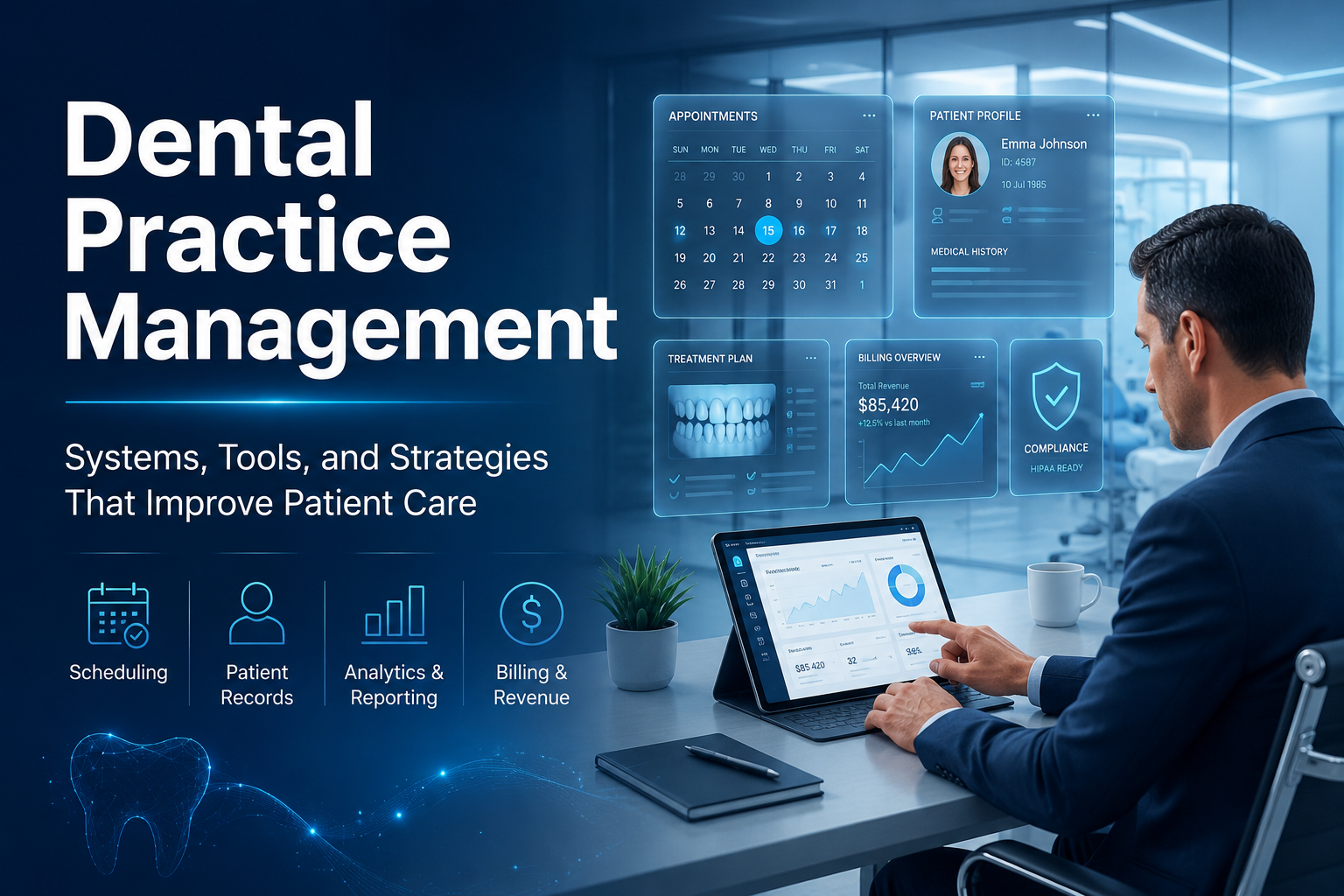
Many clinics are addressing this by adding an operational intelligence layer that makes soft tissue screening consistent and trackable: AI soft tissue screening integration, disease-wise visibility, and workflow tools (scheduling, documentation, patient follow-up) that reduce “lost to review.” scanO Engage is one example of how practices are building that structure without turning every case into a referral, because the win is not more alarms. The win is fewer untracked maybes.
The reflective question for a clinic owner is simple: when a subtle lesion appears today, does your system reliably turn it into a documented, time-bound decision, or does it depend on who saw it and how busy the chair was.
1. Why should a clinic owner be concerned about using Early Signs of oral cancer AI if oral cancer cases don’t occur often?
The risk to the business is not equal. Missing even one early lesion can cause late diagnosis, hurt the clinic’s reputation, and lead to legal trouble. Early Signs of oral cancer AI helps by ensuring screenings are more reliable and easier to track.
2. What effect does Early Signs of oral cancer AI have on chairside efficiency and daily workflows in clinics?
Standardized screenings save time since dentists can spend less effort second-guessing unclear cases and instead focus on clear plans of action. Early Signs of oral cancer AI makes documentation quicker follow-up steps simpler, and reduces unscheduled repeat checks that can disrupt the day's routine.
3. Can Early Signs of oral cancer AI lower the reliance on a clinician's personal experience?
Yes. It sets a standard for screening that applies to every patient making sure early detection remains steady no matter if the clinician is new, experienced, or during different work schedules.
4. How does Early Signs of oral cancer AI compare to traditional screening in terms of compliance and documentation?
Traditional methods often depend on quick notes and memory to review cases. Early Signs of oral cancer AI enhances tracking by connecting images to organized follow-ups. This adds to clinical documentation and makes cases easier to defend.
5. How does Tissue AI benefit a clinic owner beyond basic oral screening?
Tissue AI allows soft tissue monitoring over time helping clinic owners observe long-term changes across a group of patients. It helps in spotting recurring risk patterns and shifts their approach from handling cases as they happen to focusing on broader prevention strategies at a system level.
An AI-powered co-author focused on generating data-backed insights and linguistic clarity.
Dr. Vidhi Bhanushali is the Co-Founder and Chief Dental Surgeon at scanO . A recipient of the Pierre Fauchard International Merit Award, she is a holistic dentist who believes that everyone should have access to oral healthcare, irrespective of class and geography. She strongly believes that tele-dentistry is the way to achieve that.Dr. Vidhi has also spoken at various dental colleges, addressing the dental fraternity about dental services and innovations. She is a keen researcher and has published various papers on recent advances in dentistry.